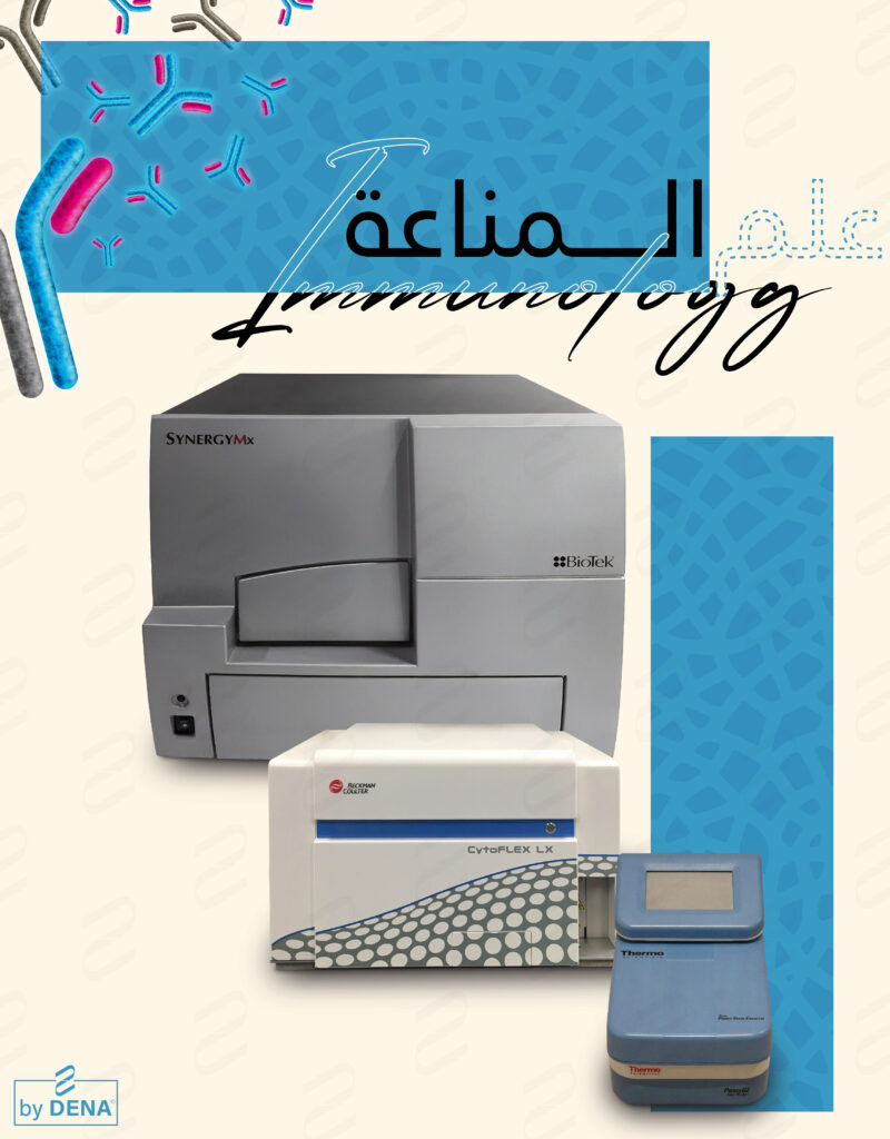
The Thermo Scientific Pierce Power Blotter is a specialized instrument designed to optimize the transfer of proteins from polyacrylamide gels (like those used in SDS-PAGE) onto membranes, a key step in the Western blotting process. Western blotting is a widely used technique for the detection and analysis of specific proteins in a sample. Here are some key aspects of the Pierce Power Blotter:
Key Features and Functions
- Efficient Protein Transfer: The Pierce Power Blotter is designed to provide rapid and efficient transfer of proteins. This is crucial for high-quality Western blots, as effective transfer ensures that the proteins are accurately represented on the membrane for subsequent detection.
- Semi-Dry Transfer Technology: Unlike traditional wet transfer systems, the Pierce Power Blotter often utilizes a semi-dry transfer method. This approach typically requires less buffer and can be faster while still maintaining high transfer efficiency.
- Uniform Electric Field: The system is engineered to provide a uniform electric field across the gel, ensuring consistent and even transfer of proteins from the gel to the membrane. This uniformity is important for the reproducibility of results.
- Compatibility with Different Gel Sizes: The Pierce Power Blotter is generally compatible with a range of gel sizes and types, allowing for flexibility in experimental design.
- Rapid Transfer Times: One of the significant advantages of this system is its ability to perform rapid protein transfers, often in a matter of minutes, compared to the hour or more required for traditional wet transfer methods.
- User-Friendly Interface: These systems typically come with an intuitive interface, making it relatively straightforward to set up and run protein transfers.
- Versatile Application: It’s suitable for transferring a wide range of protein sizes, making it a versatile tool for various Western blotting applications.
Applications in Research and Diagnostics
- The Pierce Power Blotter is commonly used in laboratories performing Western blotting for protein analysis. This can include research in molecular biology, biochemistry, and medical diagnostics.
- It is particularly valuable in settings where time efficiency and reproducibility of protein transfers are critical.
Advantages Over Traditional Methods
- Speed: Significantly faster than traditional wet transfer methods.
- Efficiency: Improved transfer efficiency, particularly for high molecular weight proteins.
- Ease of Use: Simplifies the Western blotting workflow, making it more accessible to users with varying levels of expertise.
Considerations
- Cost and Accessibility: Instruments like the Pierce Power Blotter can be an investment for some labs, so it’s important to evaluate the cost-benefit based on the lab’s specific needs.
- Compatibility with Existing Protocols: Researchers need to ensure that the system is compatible with their existing Western blotting protocols and reagents.
For detailed specifications, operational guidelines, and to ensure compatibility with specific applications, it’s best to consult the product literature provided by Thermo Fisher Scientific or directly contact their technical support. They can provide the most current information, user manuals, and technical assistance related to the Pierce Power Blotter.
Enzyme-Linked Immunosorbent Assay (ELISA) and its associated equipment, such as microplate readers, play a critical role in various fields of biological and medical sciences. ELISA is a popular analytical technique used for detecting and quantifying substances such as peptides, proteins, antibodies, and hormones. Here’s an overview of its importance and applications:
Importance of ELISA
- Sensitivity and Specificity: ELISA is highly sensitive and specific, making it a valuable tool for detecting specific analytes in complex mixtures.
- Quantitative Analysis: It allows for precise quantitative analysis of target analytes, which is crucial in both research and clinical diagnostics.
- Versatility: ELISA can be adapted to detect a wide range of molecules, making it a versatile tool in various scientific studies.
- Non-Radioactive: Unlike some older assay methods, ELISA does not require radioactive materials, making it safer and more accessible in different laboratory settings.
- High Throughput: ELISA can be performed in a microplate format, allowing simultaneous analysis of multiple samples. This high-throughput capability is essential for large-scale studies and screenings.
Applications of ELISA
- Disease Diagnosis: ELISA is widely used in medical diagnostics for the detection of various pathogens (like viruses and bacteria) and their antibodies, aiding in the diagnosis of infections such as HIV, Lyme disease, and certain types of hepatitis.
- Allergy Testing: It is employed to detect allergen-specific IgE antibodies, helping to diagnose allergies.
- Hormone and Biomarker Testing: ELISA is used to measure hormone levels in blood, which is vital in the diagnosis and monitoring of endocrine disorders and during fertility treatments.
- Drug Monitoring: It helps in therapeutic drug monitoring by quantifying drug levels in the blood to ensure optimal dosing.
- Food Industry: ELISA is used to detect potential allergens in food products, like peanuts or gluten, and to test for food contaminants.
- Research Applications: In research, ELISA is used to study protein-protein interactions, cytokine profiling, and in the development and testing of vaccines.
- Environmental Testing: It aids in the detection of environmental pollutants, such as pesticides and industrial chemicals.
Microplate Readers in ELISA
- Detection and Analysis: Microplate readers are essential for detecting the optical changes (colorimetric, fluorometric, or luminescent) in an ELISA assay and converting them into quantitative data.
- Automation and Data Processing: These devices often come with software for automated data analysis, which is crucial for interpreting ELISA results, especially in high-throughput settings.
- Flexibility: Modern microplate readers can handle different assay formats and detection methods, increasing the versatility of ELISA applications.
Last Word
ELISA and microplate readers are indispensable tools in diagnostics, research, and various industrial applications due to their high sensitivity, specificity, and adaptability. They have significantly contributed to advancements in healthcare, environmental monitoring, and scientific research.
